Research's Attempt to Objectively Assess Tinnitus

Discussion in 'Research News' started by Frédéric, Jun 21, 2017.

    1. AUTHOR
      AUTHOR
      Frédéric

      Frédéric Member Podcast Patron Benefactor Advocate

      Location:
      Marseille, France
      Tinnitus Since:
      11/19/2012
      Cause of Tinnitus:
      acoustic trauma
      A PhD dissertation that concludes that GPIAS works in humans.

      Change in Acoustic Startle as an Indicator of Continuous Tonal Tinnitus

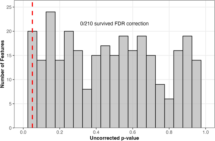
      Abstract: Currently, there is no accepted objective measure of tinnitus in humans. The gap prepulse inhibition of acoustic startle (GPIAS) paradigm is an objective measure that has been used in the animal model to identify tinnitus based on the theory of tinnitus filling in the silent gap that would normally promote startle inhibition. The current study applied the GPIAS paradigm in human subjects with normal hearing thresholds without hyperacusis. Individuals with continuous tonal tinnitus (N=31) characterized their tinnitus by adjusting a signal to match the frequency, bandwidth, and intensity. These individual parameters were used to create maximally matched background sounds in the GPIAS paradigm for each subject. A group without tinnitus (N=8) also participated using the averaged parameter values of the background sound from the group with tinnitus. Startle inhibition percentage was calculated by comparing ocular EMG blinking amplitudes between gap embedded conditions and the condition without a gap. As expected, the group with no tinnitus revealed startle inhibition as evidenced by reduced EMG blink amplitudes when the background sound was interrupted by a silent gap prior to the startle impulse (100 dB SPL white noise). The group with tinnitus did not have a significant startle inhibition in this same condition supporting the theory that the background sound carefully matched to their tinnitus eliminated the perception of a silent gap, thereby removing the cue that would produce startle inhibition. Gradually increasing the contrast between the individual’s continuous tonal tinnitus and ongoing background sound leads to a nonlinear change in startle inhibition percentage, providing guidelines for how closely the background sound needs to match the tinnitus of an individual in v order to get the expected result of no startle inhibition when tinnitus is filling in the gap. Collectively, these findings support the use of the GPIAS paradigm for objectively identifying continuous tonal tinnitus in humans. Further, certain deviations in frequency, intensity, or bandwidth in the ongoing background sound from the tinnitus match result in startle inhibition, which may help explain the inconsistent findings across human GPIAS studies and allow more confidence for animal researchers to use GPIAS for animal tinnitus studies.
       

      Attached Files:

      • Informative Informative x 1
    2. weab00
      Gloomy

      weab00 Member Benefactor

      Location:
      nunya
      Tinnitus Since:
      05/2019
      Cause of Tinnitus:
      some good mf music
      So when will this technology become mainstream in audiology offices?
       
    3. AUTHOR
      AUTHOR
      Frédéric

      Frédéric Member Podcast Patron Benefactor Advocate

      Location:
      Marseille, France
      Tinnitus Since:
      11/19/2012
      Cause of Tinnitus:
      acoustic trauma
      Well, I relay research publications even if there is no significant results.

      Auditory evoked potential P300 characteristics in adults with and without idiopathic bilateral tinnitus

      Background and Aim: Based on neurophysiological measurements, auditory and non-auditory pathways are involved in tinnitus. People who experience tinnitus may suffer from several problems such as attention disorder. The auditory evoked potential P300 is an endogenous response and depends on cognitive processes like attention. The purpose of this study was to compare the auditory P300 characteristics (amplitude and latency) in adults with and without tinnitus.

      Methods: Participants were 20 adults with idiopathic bilateral tinnitus with mean duration of 8.4 ± 4.73 months, and 20 healthy adults. The P300 was recorded using oddball paradigm consisted of two standard (1000 Hz) and target stimuli (2000 Hz). To reduce eye blink during recording, all participants was instructed to look at and fixate on a dot sign located in front of them. The tinnitus handicap inventory (THI) was completed and pitch matching (PM) and loudness matching (LM) were measured in tinnitus group.

      Results: P300 amplitude was lower at both Fz and Cz electrode placements in tinnitus patients compared to the normal group, but it was not statistically significant (p = 0.57). Tinnitus patients had delayed latency at Fz and Cz, but this difference was not significant either psychometric and psychoacoustic assessment had no statistically significant correlation with P300 amplitude and latency.

      Conclusion: It seems that P300 characteristics are not different between adults with and without idiopathic bilateral tinnitus, may be due to using low sample size.
       

      Attached Files:

    4. AUTHOR
      AUTHOR
      Frédéric

      Frédéric Member Podcast Patron Benefactor Advocate

      Location:
      Marseille, France
      Tinnitus Since:
      11/19/2012
      Cause of Tinnitus:
      acoustic trauma
      Neuroanatomical Alterations in Patients With Tinnitus Before and After Sound Therapy: A Combined VBM and SCN Study

      Background: Many neuroanatomical alterations have been detected in patients with tinnitus in previous studies. However, little is known about morphological and structural covariance network (SCN) changes before and after long-term sound therapy. This study aimed to explore alterations in brain anatomical and SCN changes in patients with idiopathic tinnitus using voxel-based morphometry (VBM) analysis 24 weeks before and after sound therapy.

      Methods: Thirty-three tinnitus patients underwent magnetic resonance imaging scans at baseline and after 24 weeks of sound therapy. Twenty-six age- and sex-matched healthy control (HC) individuals also underwent two scans over a 24-week interval; 3.0T MRI and high-resolution 3D structural images were acquired with a 3D-BRAVO pulse sequence. Structural image data preprocessing was performed using the VBM8 toolbox. The Tinnitus Handicap Inventory (THI) score was acquired in the tinnitus group to assess the severity of tinnitus and tinnitus-related distress. Two-way mixed model analysis of variance (ANOVA) and post hoc analyses were performed to determine differences between the two groups (patients and HCs) and between the two scans (at baseline and at the 24th week). Two-sample t tests, paired-samples t tests, and Pearson’s correlation analysis were used in the post hoc analysis.

      Results: Interaction effects between the two groups and the two scans demonstrated signicantly different gray matter (GM) volume in the right parahippocampus gyrus, right caudate, left superior temporal gyrus, left cuneus gyrus and right calcarine gyrus; we found signicantly decreased GM volume in the above ve brain regions among the tinnitus patients before sound therapy (baseline) compared to that in the HC group. The 24-week sound therapy group demonstrated signicantly greater brain volume compared with the baseline group among these brain regions. We did not nd signicant differences in brain regions between the 24-week sound therapy and HC groups. The SCN results showed that the left superior temporal gyrus and left rolandic operculum were signicantly different in nodal eciency, nodal degree centrality and nodal betweenness centrality after FDR correction. Decreased THI scores and GM volume changes between the left thalamus and right thalamus were not correlated.

      Conclusions: This study characterized the effect of sound therapy on brain GM volume, especially in the left superior temporal lobe. Notably, sound therapy had a normalizing effect on tinnitus patients.

      Full article: see PDF file.
       

      Attached Files:

      • Informative Informative x 1
    5. AUTHOR
      AUTHOR
      Frédéric

      Frédéric Member Podcast Patron Benefactor Advocate

      Location:
      Marseille, France
      Tinnitus Since:
      11/19/2012
      Cause of Tinnitus:
      acoustic trauma
      Otoacoustic emissions and contralateral suppression in tinnitus sufferers with normal hearing

      Abstract


      Background

      The general consensus on the role of hearing loss in generating tinnitus is not relevant in tinnitus patients with normal hearing thresholds. One source of tinnitus may be related to damage to outer hair cells (OHC) of the cochlea. If the OHC of the human cochlea are to be involved in the generation of tinnitus, testing of Otoacoustic emissions (OAE) could provide a reliable means of recording OHC dysfunction. We investigated the role of OHC and cochlear efferent system in tinnitus development in normal hearing ears through studying of Distortion Product Otoacoustic Emissions (DPOAE) and Transient Evoked Otoacoustic Emissions (TEOAE) amplitudes, contralateral suppression amplitudes and suppression value in 15 normal hearing tinnitus patients and 15 control subjects.

      Results

      Mean f2 DPOAE amplitudes and contralateral suppression were significantly lower in tinnitus group compared to controls for all frequencies from 1001 to 6348 Hz. Suppression values of DPOAEs revealed lower but not significant difference between tinnitus and control groups for all frequencies except 1587 and 6348 Hz. TEOAE amplitudes and contralateral suppression were significantly lower in tinnitus groups for all frequencies from 1000 to 4000 Hz compared to the control group. Suppression value of TEOAEs revealed no significant difference between the two groups for all frequencies except 3000 and 4000 Hz were significantly lower in the tinnitus group compared to the control group.

      Conclusions

      Normal hearing manifested by pure tone audiometry in non-vascular tinnitus sufferers does not exclude OHC and/or cochlear efferent pathology.

      Full article: https://ejo.springeropen.com/articles/10.1186/s43163-020-00030-4
       
      • Informative Informative x 1
    6. AUTHOR
      AUTHOR
      Frédéric

      Frédéric Member Podcast Patron Benefactor Advocate

      Location:
      Marseille, France
      Tinnitus Since:
      11/19/2012
      Cause of Tinnitus:
      acoustic trauma
      Effect of age on the gap-prepulse inhibition of the cortical N1-P2 complex in humans as a step towards an objective measure of tinnitus

      The gap-prepulse inhibition of the acoustic startle reflex has been widely used as a behavioral method for tinnitus screening in animal studies. The cortical-evoked potential gap-induced inhibition has also been investigated in animals as well as in human subjects. The present study aimed to investigate the effect of age on the cortical N1-P2 complex in the gap-prepulse inhibition paradigm. Fifty-seven subjects, aged 20 to 68 years, without continuous tinnitus, were tested with two effective gap conditions (embedded gap of 50- or 20-ms duration). Retest sessions were performed within one month. A significant gap-induced inhibition of the N1-P2 complex was found in both gap durations. Age differently affected the inhibition, depending on gap duration. With a 50-ms gap, the inhibition decreased significantly with the increase in age. This age-inhibition relationship was not found when using a 20-ms gap. The results were reproducible in the retest session. Our findings suggest that the interaction between age and gap duration should be considered when applying the gap-induced inhibition of the cortical-evoked potential as an objective measure of tinnitus in human subjects. Further studies with tinnitus patients are warranted to identify gap duration that would minimize the effects of age and maximize the difference in the inhibition between those with and without tinnitus.

      Full article: https://journals.plos.org/plosone/article?id=10.1371/journal.pone.0241136
       
      • Informative Informative x 1
    7. AUTHOR
      AUTHOR
      Frédéric

      Frédéric Member Podcast Patron Benefactor Advocate

      Location:
      Marseille, France
      Tinnitus Since:
      11/19/2012
      Cause of Tinnitus:
      acoustic trauma
      Within-Subject Comparisons of the Auditory Brainstem Response and Uncomfortable Loudness Levels in Ears With and Without Tinnitus in Unilateral Tinnitus Subjects With Normal Audiograms.

      Objective: To evaluate whether cochlear synaptopathy is a common pathophysiologic cause of tinnitus in individuals with normal audiograms.

      Study design: Prospective study.

      Setting: Tertiary referral center.

      Methods: We enrolled 27 subjects with unilateral tinnitus and normal symmetric hearing thresholds, and 27 age- and sex-matched control subjects with normal symmetric hearing thresholds. We measured 1) the amplitudes of waves I and V with 90 dB nHL click stimuli in quiet conditions; 2) the latency shift of wave V with 80 dB nHL click stimuli in background noise, varying from 40 dB HL to 70 dB HL; and 3) uncomfortable loudness levels (UCLs) at 500 Hz and 3000 Hz pure tones.

      Results: There were no significant differences in the wave V/I amplitude ratio or the latency shift in wave V with increasing noise levels among the tinnitus ears (TEs), nontinnitus ears (NTEs), and control ears. There were no significant differences in UCLs at 500 Hz or 3000 Hz between TEs and NTEs, but the UCLs were lower in TEs (mean 111.3 dB or 104.1 dB) and NTEs (mean 109.4 dB or 100.6 dB) than in control ears (mean 117.9 dB or 114.1 dB, p < 0.017). No subject met our criteria for cochlear synaptopathy or increased central gain in terms of all three parameters.

      Conclusion: Based on these results for UCL, increased central gain is a major mechanism of tinnitus in humans with normal audiograms. However, this compensatory mechanism for reduced auditory input may originate from other pathophysiologic factors rather than from cochlear synaptopathy.

      Source: https://europepmc.org/article/med/33177407
       
      • Informative Informative x 1
    8. AUTHOR
      AUTHOR
      Frédéric

      Frédéric Member Podcast Patron Benefactor Advocate

      Location:
      Marseille, France
      Tinnitus Since:
      11/19/2012
      Cause of Tinnitus:
      acoustic trauma
      Correlation Analysis of EEG Brain Network with Modulated Acoustic Stimulation for Chronic Tinnitus Patients

      The acoustic stimulation influences of the brain is still unveiled, especially from the brain network point, which can reveal how interaction is propagated and integrated between different brain zones for chronic tinnitus patients. We specifically designed a paradigm to record the electroencephalograms (EEGs) for tinnitus patients when they were treated with consecutive acoustic stimulation neuromodulation therapy for up to 75 days, using the tinnitus handicap inventory (THI) to evaluate the tinnitus severity or the acoustic stimulation treatment efficacy, and the EEG to record the brain activities every 2 weeks. Then, we used an EEG-based coherence analysis to investigate if the changes in brain network consistent with the clinical outcomes can be observed during 75-days acoustic treatment. Finally, correlation analysis was conducted to study potential relationships between network properties and tinnitus handicap inventory score change. The EEG network became significantly weaker after long-term periodic acoustic stimulation treatment, and tinnitus handicap inventory score changes or the acoustic stimulation treatment efficacy are strongly correlated with the varying brain network properties. Long-term acoustic stimulation neuromodulation intervention can improve the rehabilitation of chronic tinnitus patients, and the EEG network provides a relatively reliable and quantitative analysis approach for objective evaluation of tinnitus clinical diagnosis and treatment.

      Source: https://pubmed.ncbi.nlm.nih.gov/33216716/
       
      • Informative Informative x 1
    9. AUTHOR
      AUTHOR
      Frédéric

      Frédéric Member Podcast Patron Benefactor Advocate

      Location:
      Marseille, France
      Tinnitus Since:
      11/19/2012
      Cause of Tinnitus:
      acoustic trauma
      Altered effective brain network topology in tinnitus: An EEG source connectivity analysis

      Tinnitus is defined as the auditory phantom perception in the absence of any objective external sound source. In this paper, we used the resting-state electroencephalography (EEG) data to reconstruct the neural sources based on the unit-noise-gain linearly constrained minimum-variance (LCMV) beamformer and applied the effective connectivity analysis on the reconstructed sources to examine the directional neuronal interactions between brain regions in tinnitus patients compared to the healthy controls. We found significantly disrupted patterns of effective connectivity in several brain areas including the frontal, temporal, and occipital cortices as well as the caudate nucleus. Particularly, significant aberrant causal couplings were observed in the orbitofrontal cortex, inferior frontal gyrus_triangular, and parahippocampal region that could potentially illustrate the auditory information retrieval, perception, and evaluation of the phantom sound in the brain of tinnitus patients. Furthermore, topological alterations of the brain network were investigated using graph theoretical analysis. Our findings demonstrated significantly decreased both global integration and segregation of the brain network in tinnitus patients accompanied by the topological shift of tinnitus network to a more random structure in the high-frequency bands. These findings were consistent with the hypothesis of the brain network deviation from small-worldness topology accompanied by reduced global integration in brain-related disorders.

      https://www.sciencedirect.com/science/article/abs/pii/S1746809420304468
       
    10. AUTHOR
      AUTHOR
      Frédéric

      Frédéric Member Podcast Patron Benefactor Advocate

      Location:
      Marseille, France
      Tinnitus Since:
      11/19/2012
      Cause of Tinnitus:
      acoustic trauma
      Pity we don't know how these tinnitus sufferers can switch on/off their tinnitus.

      Switching Tinnitus-On: Maps and Source Localization of Spontaneous EEG

      Objective
      To identify the spectrotemporal changes and sources in patients that could “turn on” tinnitus with multichannel electroencephalography (EEG) system.

      Methods
      Multichannel EEG was recorded from six patients during the Tinnitus-On and Tinnitus-Off states. The EEG power spectrum and eLORETA-based sources were measured.

      Results
      There was a global increase in delta and theta during Tinnitus-On plus large changes in alpha 1 and alpha 2. During the Tinnitus-On state, many new sources in delta, theta, alpha 1 and gamma bands emerged in the opposite hemisphere in the inferior temporal gyrus (Brodmann area, BA 20), middle temporal gyrus (BA 21), lateral perirhinal cortex (BA 36), ventral entorhinal cortex (BA 28) and anterior pole of the temporal gyrus (BA 38).

      Conclusions
      The emergence of new delta, theta and gamma band sources in the inferior temporal gyrus (BA 20), middle temporal gyrus (BA 21) and lateral perirhinal cortex (BA 36) plus the appearance of new delta and theta sources in the ventral entorhinal cortex (BA28) and anterior pole of the temporal lobe (BA 38) may comprise a network capable of evoking the phantom sound of tinnitus by simultaneously engaging brain regions involved in memory, sound recognition, and distress which together contribute to tinnitus severity.

      Significance
      The sudden appearance of new sources of activity in the opposite hemisphere within the inferior temporal gyrus, middle temporal gyrus and perirhinal cortex may initiate the perception of tinnitus perception.

      Source: https://www.sciencedirect.com/science/article/abs/pii/S1388245720305575
       
      • Informative Informative x 2
    11. AUTHOR
      AUTHOR
      Frédéric

      Frédéric Member Podcast Patron Benefactor Advocate

      Location:
      Marseille, France
      Tinnitus Since:
      11/19/2012
      Cause of Tinnitus:
      acoustic trauma
      An article on trends in otologic research:

      Clinical trials in otology: Examining trends and framework for prioritization

      Results
      There were 992 otology clinical trials from 2008 to 2018.457 (46.1%) were completed and 94 (9.5%) were discontinued. Industry remained the highest (76.5%) contributor to otology clinical trials. The otologic conditions studied, from most common to least common, include hearing loss (40.6%), vestibulopathy (18.8%), tinnitus (18.8%), and otitis media (15.1%). The number of otology clinical trials increased by an average of 12.0 trials per year from 2008 to 2018 (p < 0.001). The number of otology clinical trials focusing on hearing loss and vestibulopathy significantly increased over the studied period (p < 0.001), while those focusing on tinnitus and otitis media did not (p = 0.09 and p = 0.20, respectively). The majority of clinical trials on each of these four conditions focused on treatment options.

      Conclusion
      Our study describes trends in otology clinical trials registered on clinicaltrials.gov from 2008 through 2018. The total number of clinical trials over this time period increased significantly, driven by trials investigating hearing loss and vestibulopathy. Furthermore, most clinical trials were industry-sponsored and focused on treatment modalities. Our study provides an outline of otology clinical trials registered in a US web-based database, which may be of use for the development of future clinical trials.
       
      • Informative Informative x 1
    12. AUTHOR
      AUTHOR
      Frédéric

      Frédéric Member Podcast Patron Benefactor Advocate

      Location:
      Marseille, France
      Tinnitus Since:
      11/19/2012
      Cause of Tinnitus:
      acoustic trauma
      From the same team, investigation is enhanced via probes placed in the auditory canal.

      Tinnitus and auditory cortex; Using adapted functional near‐infrared‐spectroscopy to expand brain imaging in humans

      Objectives
      Phantom sound perception (tinnitus) may arise from altered brain activity within auditory cortex. Auditory cortex neurons in tinnitus animal models show increased spontaneous firing rates. This may be a core characteristic of tinnitus. Functional near‐infrared spectroscopy (fNIRS) has shown similar findings in human auditory cortex. Current fNIRS approaches with cap recordings are limited to ∼3 cm depth of signal penetration due to the skull thickness. To address this limitation, we present an innovative fNIRS approach via probes adapted to the external auditory canal. The adapted probes were placed deeper and closer to temporal lobe of the brain to bypass confining skull bone and improve neural recordings.

      Methods
      Twenty adults with tinnitus and 20 nontinnitus controls listened to periods of silence and broadband noise (BBN) during standard cap and adapted ear canal fNIRS neuroimaging. The evaluators were not blinded, but the protocol and postprocessing for the two groups were identical.

      Results
      Standard fNIRS measurements in participants with tinnitus revealed increased auditory cortex activity during silence that was suppressed during auditory stimulation with BBN. Conversely, controls displayed increased activation with noise but not during silence. Importantly, adapted ear canal fNIRs probes showed similar hemodynamic responses seen with cap probes in both tinnitus and controls.

      Conclusions
      In this proof of concept study, we have successfully fabricated, adapted, and utilized a novel fNIRS technology that replicates established findings from traditional cap fNIRS probes. This exciting new innovation, validated by replicating previous and current cap findings in auditory cortex, may have applications to future studies to investigate brain changes not only in tinnitus but in other pathologic states that may involve the temporal lobe and surrounding brain regions.
       
      • Winner Winner x 1
      • Informative Informative x 1
Loading...

Share This Page